
Authentic images only
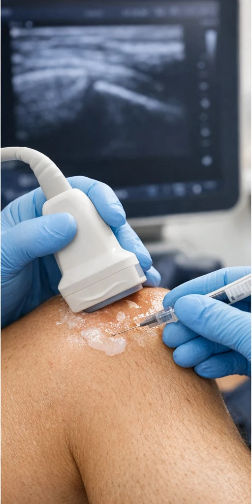
The clinical atlas includes original ultrasound teaching examples alongside procedural and educational illustrations. Images should be interpreted in clinical context. This distinction is deliberate: a photograph of a scanner or procedure must not be described as a diagnostic scan showing a specific pathology.
Requirements for each clinical image
- Original still or cine image obtained with appropriate consent and rights.
- Complete removal of identifiers and metadata that could identify a patient.
- Confirmed region, side, probe orientation and scanning plane.
- Labelled anatomical structures and findings reviewed by the scanning clinician.
- Short plain-language explanation alongside detailed clinician interpretation.
- Limits of diagnostic confidence, alternatives and clinical correlation.
- Relevant and individually verified references.
Ultrasound teaching examples
Priority examples: shoulder supraspinatus tendon, subacromial-subdeltoid bursa, knee suprapatellar effusion, Achilles tendon, plantar fascia, common extensor origin at the elbow, greater trochanteric region, and wrist flexor tendons. No scan will be portrayed as a real patient case until the original image is supplied and clinically checked.
Available image in this prototype
This information is for general education and does not replace advice from a clinician who has assessed you.