
Posterior elbow and triceps insertion
A clinician-guided explanation of a real elbow ultrasound image, its anatomy, and how it fits into assessment.
All real scan examples · Elbow ultrasound atlas
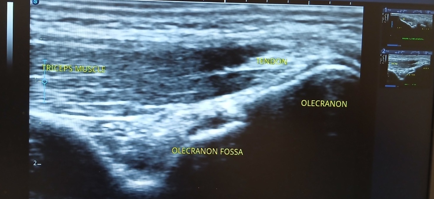
Supplied ultrasound screen photograph, presented without artificial reconstruction. Not a complete clinical case report.
Teaching interpretation
The image demonstrates the posterior elbow sonographic landmarks relevant to distal triceps examination and adjacent superficial structures. It is an anatomical illustration from an actual scan, not a confirmed tendon injury.
How it may relate to physiotherapy
Posterior elbow pain may arise from tendon loading, local bursitis, injury or other causes. The appropriate work-up follows history, physical examination and the exact clinical question.
Important limitations
Ultrasound may assist assessment of the distal triceps tendon and superficial fluid, but does not diagnose all causes of elbow pain.
Related care and references
Explore physiotherapy, diagnostic ultrasound and the wider atlas. These materials are educational and do not establish a diagnosis for an individual patient.