
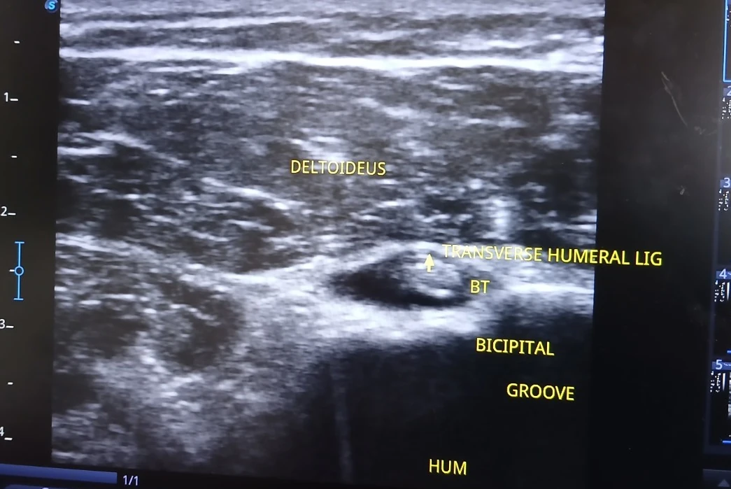
Shoulder ultrasound
Rotator cuff tendons, long-head biceps tendon, subacromial-subdeltoid bursa and calcific deposits.
Original clinician-supplied image
Read clinical guide →Home / Ultrasound atlas
Learn what ultrasound can show in six major body regions, how findings are interpreted and why imaging should follow the clinical question — not replace physiotherapy assessment.
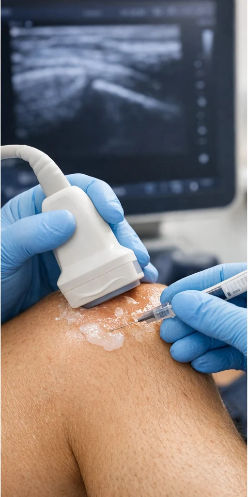
See the suprapatellar recess, popliteal Doppler, biceps groove, patellar tendon insertion and AC joint. Clear explanations accompany each clinician-supplied image.
Rotator cuff tendons, long-head biceps tendon, subacromial-subdeltoid bursa and calcific deposits.
Original clinician-supplied image
Read clinical guide →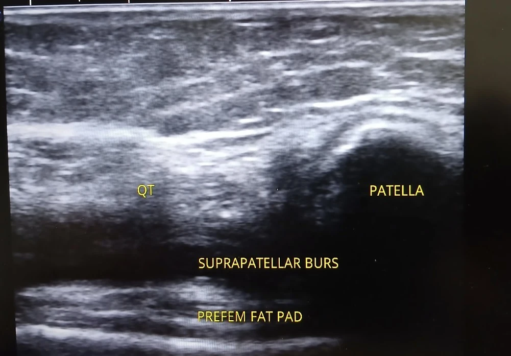
Effusion, synovial recesses, superficial tendons, bursae and some peripheral soft tissues.
Original clinician-supplied image
Read clinical guide →Achilles, peroneal and posterior tibial tendons, selected ligament structures and plantar fascia.
Read clinical guide →Common extensor tendon, flexor-pronator origin, distal biceps and olecranon bursa.
Read clinical guide →Gluteal tendons, trochanteric bursae, anterior hip joint recess and selected soft tissues.
Read clinical guide →Flexor and extensor tendons, tenosynovitis, accessible ganglia, superficial soft tissue and selected nerves.
Read clinical guide →Each guide explains relevant structures, appropriate indications, limitations, safety and how results relate to exercise rehabilitation and other treatment options. The atlas is educational and does not provide a diagnosis without clinical assessment.
Initial source register for this atlas:
This next-stage library uses actual supplied scan images rather than only generic schematics. Each page explains what the image demonstrates and where the image fits within clinical reasoning.
Browse real ultrasound photos and individual teaching guides →