
Key anatomy and structures
Gluteal tendons, trochanteric bursae, anterior hip joint recess and selected soft tissues.
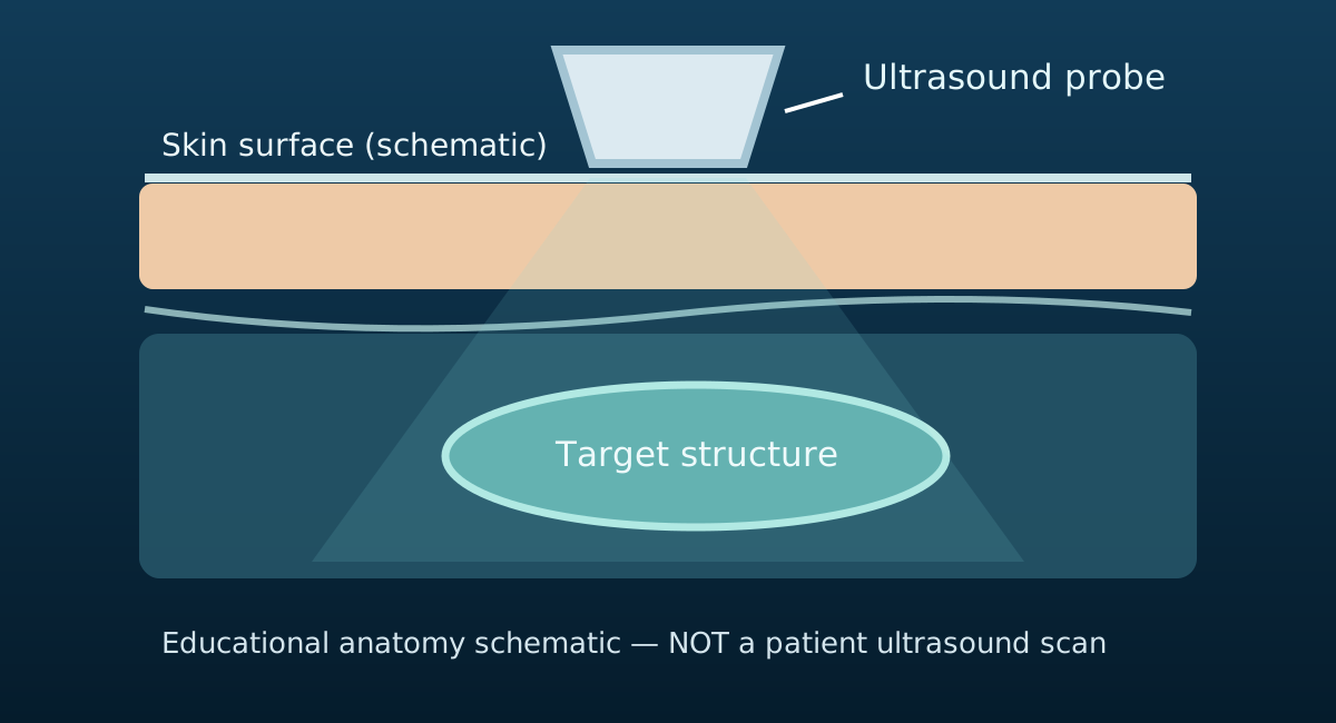
Conceptual diagram of a transducer, skin and target structure. It illustrates the scanning principle rather than the anatomy or pathology of this particular region.
When a scan might be considered
A person with lateral hip pain or groin symptoms may benefit from ultrasound when the examination identifies a focused question and the answer is likely to affect management. Scanning is not automatically required before starting physiotherapy.
Typical questions and useful findings
Gluteal tendons, trochanteric bursae, anterior hip joint recess and selected soft tissues. The clinician may examine symmetry, tissue continuity, fluid, tendon motion or a particular symptomatic area depending on the clinical indication. A finding should be reported with the exact structure, side, limitations and appropriate differential diagnosis.
Limits and clinical correlation
Some periarticular tendons are accessible. Deep hip labral pathology or extensive intra-articular assessment may require other imaging. The imaging result should be compared with the patient's history, examination, symptoms and functional goals. Incidental abnormalities can occur and do not automatically require intervention.
How this links to physiotherapy
Treatment decisions should account for strength, movement, loading tolerance, differential diagnosis and whether a scan result is likely to change the plan.
Recommended clinical pathway
1. Assess: history, functional goals, red flags and physical examination.
2. Decide: would scanning change the diagnosis, referral decision or treatment?
3. Interpret: explain findings in context and record uncertainty.
4. Treat: develop a patient-specific management plan and review progress.
Related resources
Relevant condition information · Physiotherapy assessment · Diagnostic ultrasound service.
Research and references
- NHS (n.d.) Ultrasound scan. Available at: NHS ultrasound scan (Accessed: 29 October 2026).
- NICE (2022) Osteoarthritis in over 16s: diagnosis and management. NG226. Available at: NICE NG226 recommendations (Accessed: 29 October 2026).
Assessment before imaging
The right starting point is an individual clinical assessment. A scan may be offered when appropriate and available at your location.
Contact the clinic