
Sesamoids and plantar plate: ultrasound teaching image
A clinician-guided explanation of a real foot ultrasound image, its anatomy, and how it fits into assessment.
All real scan examples · Foot ultrasound atlas
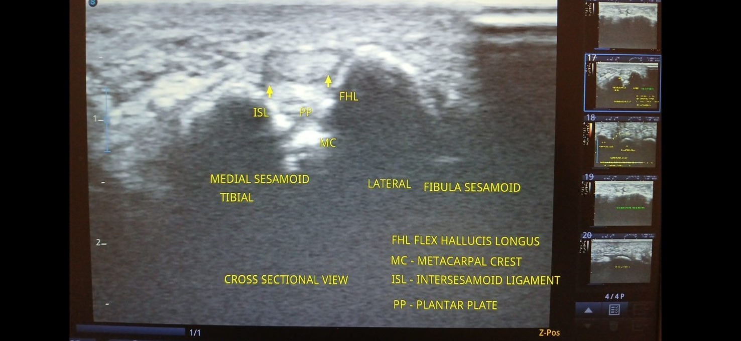
Supplied ultrasound screen photograph, presented without artificial reconstruction. Not a complete clinical case report.
Teaching interpretation
This example illustrates the relationship between the sesamoids, soft tissues and plantar aspect of the first metatarsophalangeal region. The photograph includes labels added on the source device. The source label “MC - metacarpal crest” appears anatomically inconsistent for a foot image; it requires clinician verification and has not been adopted here.
How it may relate to physiotherapy
Pain beneath the big toe may have several explanations, including local sesamoid overload, joint conditions and tendon-related pain. Imaging findings need correlation with tenderness, load response and clinical examination.
Important limitations
Foot pain can sometimes require radiography, MRI or other assessment depending on suspected fracture, osteonecrosis or intra-articular pathology. Do not infer these diagnoses from this photograph.
Related care and references
Explore physiotherapy, diagnostic ultrasound and the wider atlas. These materials are educational and do not establish a diagnosis for an individual patient.