
Power Doppler and ultrasound-guided knee procedure
A clinician-guided explanation of a real knee ultrasound image, its anatomy, and how it fits into assessment.
All real scan examples · Knee ultrasound atlas
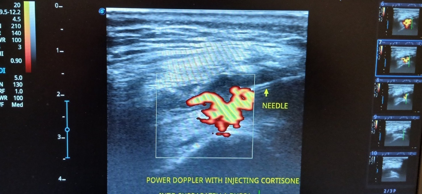
Supplied ultrasound screen photograph, presented without artificial reconstruction. Not a complete clinical case report.
Teaching interpretation
It is an example of using live imaging to assist anatomical localisation and procedural planning. The colourful Doppler signal represents blood flow information, but does not prove a particular diagnosis on its own.
How it may relate to physiotherapy
For treatment decisions, the clinician must consider indications, diagnosis, medication, patient-specific risk, consent and whether rehabilitation or other options may offer better value.
Important limitations
The photograph should not be interpreted as proof of treatment efficacy or safety; procedural accuracy and patient outcome are different questions.
Related care and references
Explore physiotherapy, diagnostic ultrasound and the wider atlas. These materials are educational and do not establish a diagnosis for an individual patient.