
Anterior knee: patella, tendon and Hoffa fat pad
A clinician-guided explanation of a real knee ultrasound image, its anatomy, and how it fits into assessment.
All real scan examples · Knee ultrasound atlas
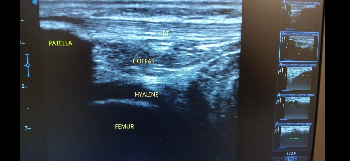
Supplied ultrasound screen photograph, presented without artificial reconstruction. Not a complete clinical case report.
Teaching interpretation
These landmarks help explain anterior knee anatomy and how soft tissues relate to the patellofemoral joint. The image does not, by itself, confirm patellofemoral pain, tendinopathy or cartilage damage.
How it may relate to physiotherapy
Anterior knee pain is often assessed through symptom history, functional loading tasks and examination. Imaging is selected only if it is likely to affect management.
Important limitations
A static sonogram cannot establish the full cause of persistent anterior knee pain; MRI or other assessment may be necessary in selected cases.
Related care and references
Explore physiotherapy, diagnostic ultrasound and the wider atlas. These materials are educational and do not establish a diagnosis for an individual patient.