
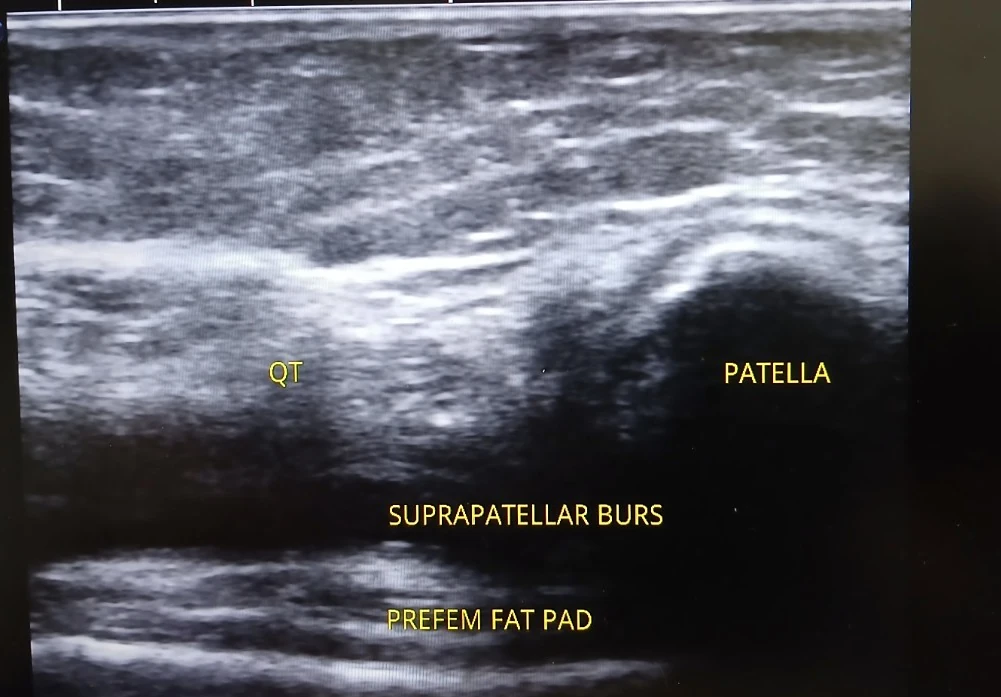
What is labelled in this image?
- Quadriceps tendon (QT)
- Patella
- Suprapatellar bursal / recess region
- Prefemoral fat pad
Why this view can be useful
The suprapatellar recess communicates with the knee joint and can be assessed for fluid. Any suspected effusion requires correlation with examination and the full scan.
What this scan does not tell us on its own
A single still cannot quantify an effusion reliably or establish the cause of knee swelling.
An ultrasound screenshot is one frame from an examination. Appropriate clinical conclusions need the history, examination, dynamic imaging and any relevant other investigations.