
Knee suprapatellar recess and effusion anatomy
A clinician-guided explanation of a real knee ultrasound image, its anatomy, and how it fits into assessment.
All real scan examples · Knee ultrasound atlas
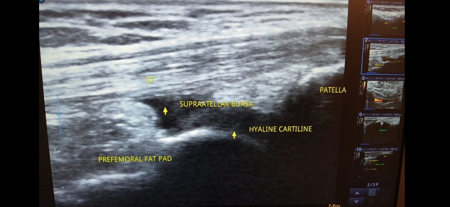
Supplied ultrasound screen photograph, presented without artificial reconstruction. Not a complete clinical case report.
Teaching interpretation
The anechoic region shown by the source labels provides a teaching example for discussing the suprapatellar recess and potential fluid. The volume or cause of any effusion cannot be established from the photograph alone.
How it may relate to physiotherapy
Knee swelling can occur after injury or alongside degenerative, inflammatory or infective conditions. A hot, acutely swollen joint or systemic illness needs prompt medical assessment.
Important limitations
Ultrasound can help identify and characterise some superficial fluid collections but does not replace clinical assessment, radiography when indicated, or aspiration for suspected infection.
Related care and references
Explore physiotherapy, diagnostic ultrasound and the wider atlas. These materials are educational and do not establish a diagnosis for an individual patient.