
Medial ankle and tibial landmark Doppler image
A clinician-guided explanation of a real ankle ultrasound image, its anatomy, and how it fits into assessment.
All real scan examples · Ankle ultrasound atlas
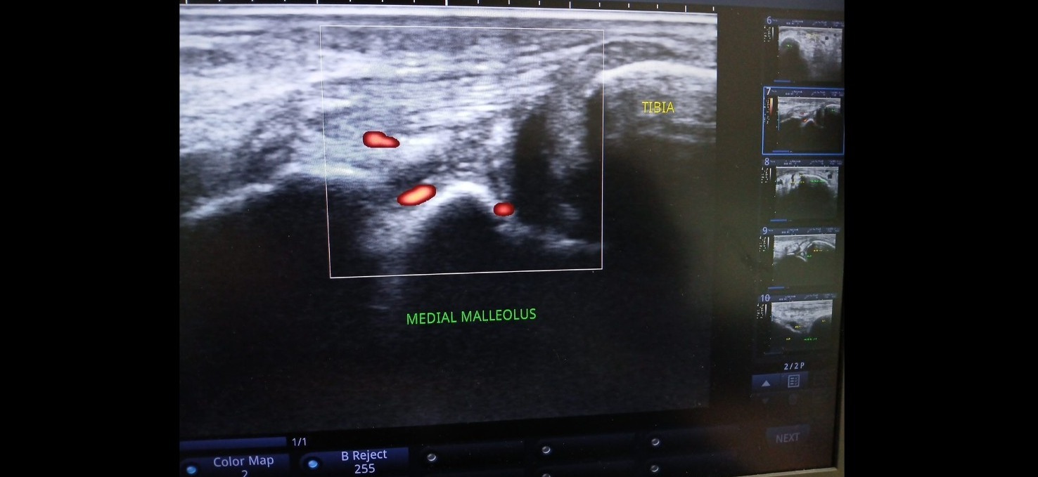
Supplied ultrasound screen photograph, presented without artificial reconstruction. Not a complete clinical case report.
Teaching interpretation
This example helps illustrate medial ankle anatomy and the use of Doppler imaging around superficial structures. The exact tendon or inflammatory diagnosis requires clinical confirmation.
How it may relate to physiotherapy
Medial ankle symptoms may involve tendon, ligament, joint or neurovascular structures. Sonography should follow a focused clinical question and examination.
Important limitations
Colour signals alone do not establish disease severity. Interpretation depends on settings, acquisition plane, symptoms and relevant clinical findings.
Related care and references
Explore physiotherapy, diagnostic ultrasound and the wider atlas. These materials are educational and do not establish a diagnosis for an individual patient.