
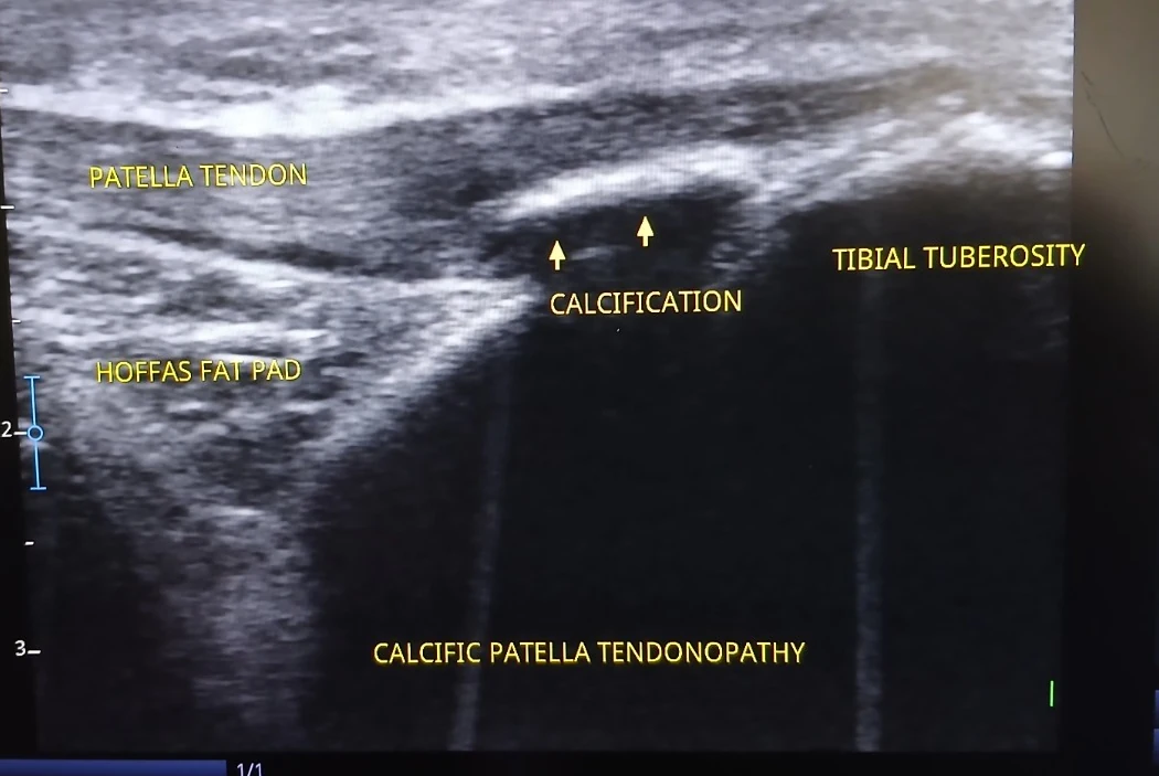
What is labelled in this image?
- Patellar tendon
- Tibial tuberosity
- Hoffa’s fat pad
- Area labelled calcification
Why this view can be useful
Tendon echotexture and the enthesis can be reviewed for changes, including calcification. The supplied annotation calls this calcific patellar tendinopathy.
What this scan does not tell us on its own
The image is a teaching example only: symptoms, palpation, loading response and other views determine whether the finding is clinically relevant.
An ultrasound screenshot is one frame from an examination. Appropriate clinical conclusions need the history, examination, dynamic imaging and any relevant other investigations.