
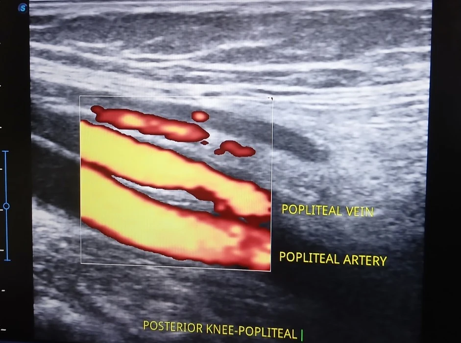
What is labelled in this image?
- Popliteal vein (labelled)
- Popliteal artery (labelled)
- Posterior knee soft tissues
- Doppler colour signal
Why this view can be useful
Doppler helps localise blood flow and adjacent vessels during focused MSK examinations, particularly when planning an intervention around the popliteal region.
What this scan does not tell us on its own
A still image does not assess venous compressibility, rule out thrombosis or demonstrate vascular health. Suspected DVT requires a formal urgent clinical pathway, not interpretation of this teaching image.
An ultrasound screenshot is one frame from an examination. Appropriate clinical conclusions need the history, examination, dynamic imaging and any relevant other investigations.