
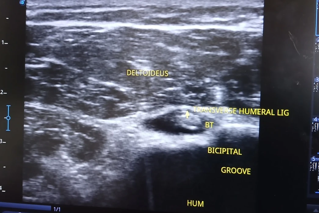
What is labelled in this image?
- Deltoid muscle
- Long-head biceps tendon (BT)
- Bicipital groove and humerus
- Transverse humeral ligament (as labelled)
Why this view can be useful
The short-axis view is one component of assessing the long head of biceps tendon position, surrounding fluid and local tendon appearance.
What this scan does not tell us on its own
Dynamic assessment, alternative views and the clinical examination are needed before interpreting suspected instability or tendon pathology.
An ultrasound screenshot is one frame from an examination. Appropriate clinical conclusions need the history, examination, dynamic imaging and any relevant other investigations.